
Fig. 5
- ID
- ZDB-FIG-110112-5
- Publication
- Robert-Moreno et al., 2010 - Characterization of new otic enhancers of the pou3f4 gene reveal distinct signaling pathway regulation and spatio-temporal patterns
- Other Figures
- All Figure Page
- Back to All Figure Page
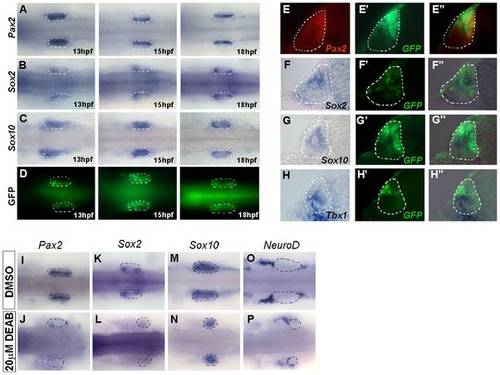
Co-localization of Pax2 and Sox2 with GFP driven by the HCNR 81675 enhancer. (A–D) Dorsal view of transgenic embryos assayed by ISH for the expression of pax2a (A), sox2 (B), sox10 (C) at 13, 15 and 18 hpf. GFP (D) displays a similar pattern than sox2 and pax2a at 15 hpf (compare A and B with D). Orientation is anterior to the left. (E–E") Double immunostaining with anti-Pax2 (E) and anti-GFP antibody (E′) in transverse sections of 15 hpf otic vesicles revealed co-localization of both proteins (E"). (F–F") GFP protein (F′) also co-localizes with sox2 mRNA (F") but not sox10 or tbx1 mRNA (G" and H"). (I–P) pax2a and sox2 expression is abolished in retinoic acid treated HCNR 81675 embryos (compare J and L to I and K, respectively) but not other genes such as sox10 or neuroD (compare N and P to M and O, respectively). Dorsal view, orientation is anterior to the left. |
| Genes: | |
|---|---|
| Antibody: | |
| Fish: | |
| Condition: | |
| Anatomical Terms: | |
| Stage Range: | 5-9 somites to 14-19 somites |