
Fig. 11
- ID
- ZDB-FIG-110112-25
- Publication
- Lin et al., 2010 - Loss of Cofilin 1 Disturbs Actin Dynamics, Adhesion between Enveloping and Deep Cell Layers and Cell Movements during Gastrulation in Zebrafish
- Other Figures
- All Figure Page
- Back to All Figure Page
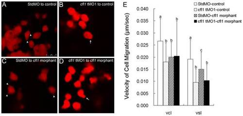
Cfl1 is required cell-autonomously for proper epiboly cell migration. Labeled cells from donor embryos injected with rhodamine-dextran in combined with StdMO (A, C) or cfl1 tMO1 (B, D) were transplanted into control embryos (A, B) and cfl1 morphants (C, D) respectively, and recorded under a confocal microscope. Pseudopods (arrowheads) and polygonal cell shape were observed in StdMO-injected cells transplanted in both control hosts (A) and cfl1 morphants (C), while blebbing-like structure (arrows) and rounded cell shape were observed in cfl1 tMO1-injected cells transplanted in both control hosts (B) and cfl1 morphants (D). The photographs shown are representative of at least 10 embryos in each experiment. Migration curvilinear velocity (Vcl) and straight line velocity (Vsl) of these StdMO- and cfl1 tMO1-injected cells in control hosts or cfl1 morphants were recorded by time-lapse epifluorescent microscopy and analyzed by SimplePCI software, respectively (E). Values between groups were compared using unpaired Student′s t-test, and those showing a significant (* p<0.05) difference are denoted by different letters. |