
Fig. 5
- ID
- ZDB-FIG-101213-16
- Publication
- Myhre et al., 2010 - Cellular differentiation in primary cell cultures from single zebrafish embryos as a model for the study of myogenesis
- Other Figures
- All Figure Page
- Back to All Figure Page
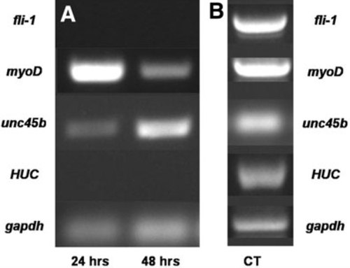
Expression of markers for endothelial, neuronal, and myogenic differentiation in ZEB cultures. ZEB cultures derived from multiple WT embryos were established and collected at 24-h intervals to obtain RNA for analysis by RTpolymerase chain reaction (PCR). (A) Detection by RT-PCR of endothelial (top row), myogenic (second and third row), and neuronal (fourth row) marker transcript expression. No fli1 (endothelial) or HUC (neuronal) marker transcription was detected in optimized ZEB cultures, whereas myoD and unc45b (muscle) transcripts were detected throughout the 2 days of culture. gapdh (fifth row) served as a positive control. (B) Control RT-PCR used bulk RNA prepared from combined embryos from 4 h to 3 days postfertilization. |