
Fig. 2
- ID
- ZDB-FIG-101213-13
- Publication
- Myhre et al., 2010 - Cellular differentiation in primary cell cultures from single zebrafish embryos as a model for the study of myogenesis
- Other Figures
- All Figure Page
- Back to All Figure Page
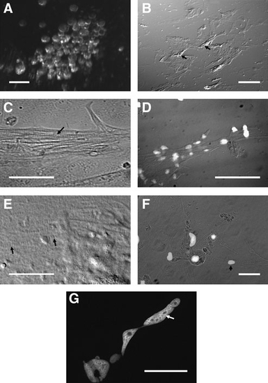
Phenotypic characterization of seZEB cultures. Single-embryo zebrafish cell cultures were established on eight-well chamber slides. (A) Live embryonic blastomeres from a single zebrafish embryo at the time of initial plating, imaged using a stereo dissecting microscope (scale bar = 0.1 mm). (B) Differential interference contrast microscopy of a single-embryo culture at low magnification (40x). Scale bar = 1mm. Arrows indicate piled-up regions of differentiating myocytes. (C–G) Differential interference contrast microscopy of differentiated myocytes in single-embryo cultures. Nuclei are indicated by DAPI staining in D and F (scale bars = 0.1mm). Arrows highlight the periodic banding pattern characteristic of striated muscle cells in panels C and E (scale bars = 0.05 mm). Arrowhead in F indicates an isolated, single-nucleated myocyte. (G) DAPI-stained single myocyte, demonstrating multiple and elongated nuclei (arrow). Scale bar = 0.05mm. |