
Fig. 1
- ID
- ZDB-FIG-101213-12
- Publication
- Myhre et al., 2010 - Cellular differentiation in primary cell cultures from single zebrafish embryos as a model for the study of myogenesis
- Other Figures
- All Figure Page
- Back to All Figure Page
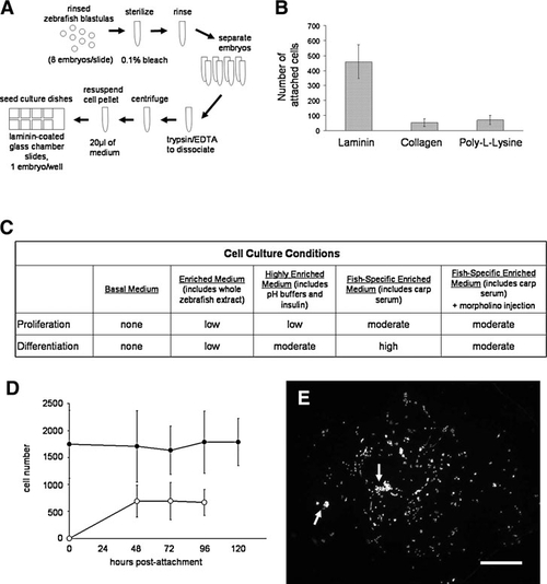
Optimal substrates and media for single-embryo zebrafish embryonic blastomere (seZEB) culture. (A) Schematic depiction of the single-embryo culture method, from embryo collection to seeding of culture slides. (B) Substrates were tested for optimal cell attachment and subsequent differentiation of ZEB cultures, including 25 μg/mL laminin, bulk rat-tail collagen, and 0.1mg/mL poly-L-lysine, as assessed by total cell count after 24 h of culture (n=24 single-embryo cultures per treatment). (C) Cell culture media were tested to determine qualitative effects on proliferation and cellular differentiation. Cell proliferation was assessed by total cell count after 4 days of culture, and differentiation was assessed by the presence or absence of morphologically distinguishable myocytes. Relative density of myocytes indicated low, medium, or high rate of differentiation (n=40–64 cultures examined per treatment). (D) Cell proliferation in highly enriched media was sufficient to overcome initial cell death after bleaching of embryos, as determined by counts of adherent cells after DAPI staining (black circles). The percentage of differentiated cells was determined by actin costaining (open circles), n=24 cultures. (E) A representative spot culture stained with DAPI to show the distribution of cell nuclei after differentiation. Arrows indicate clusters of differentiated myocytes. Scale bar=1 mm. |