
FIGURE
Fig. S1
Fig. S1
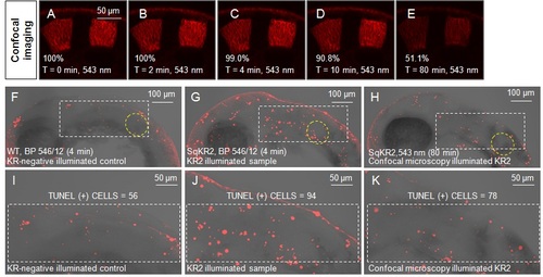
Illumination by the confocal microscope laser is inefficient in causing apoptosis. (A-E) Changes in fluorescence intensity of SqKR2 embryo during 80 minutes of continuous confocal imaging. (F-K) Illumination by green light of mercury lamp in the widefield mode increases apoptosis in the hindbrain of the SqKR2 embryo (G, J). Relatively few apoptotic cells in the SqKR2 embryo were detected following continuous confocal imaging (H, K). The otic vesicle is defined by yellow broken line (Figure 2F-H). |
Expression Data
Expression Detail
Antibody Labeling
Phenotype Data
Phenotype Detail
Acknowledgments
This image is the copyrighted work of the attributed author or publisher, and
ZFIN has permission only to display this image to its users.
Additional permissions should be obtained from the applicable author or publisher of the image.
Full text @ BMC Dev. Biol.