
Fig. 7
- ID
- ZDB-FIG-101111-7
- Publication
- Perälä et al., 2010 - Conservation, expression, and knockdown of zebrafish plxnb2a and plxnb2b
- Other Figures
- All Figure Page
- Back to All Figure Page
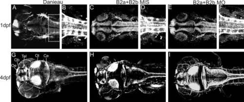
Whole-mount staining with anti-acetylated α-tubulin antibody to view axon projections in 1 days postfertilization (dpf) and 4 dpf zebrafish. A-F: Confocal scans of 1dpf zebrafish embryos. A: A dorsal view of the head of a Danieau-treated embryo. B: A ventral view of the spinal axons (area boxed in A). C-F: B2aMIS+B2bMIS-treated (5 ng+5 ng, C,D) and B2aMO+B2BMO-treated (5 ng+5 ng, E,F) embryos. G–I: Ventral stacks of 4 dpf zebrafish embryo head after Danieau-treatment (G), B2a+B2bMIS treatment (H), and B2aMO+B2bMO treatment (I). Ce, cerebellum; Ob, olfactory bulb; Oe, olfactory epithelium; Ot, optic tectum; Tel, telencephalon. |