
Fig. 4
- ID
- ZDB-FIG-101111-18
- Publication
- Barresi et al., 2010 - Essential genes for astroglial development and axon pathfinding during zebrafish embryogenesis
- Other Figures
- All Figure Page
- Back to All Figure Page
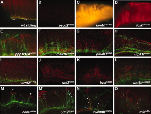
Hindbrain mutants. A-O: Lateral views of hindbrains of (A) wild-type and (B-O) mutant embryos. A: Brackets show the segmental organization of radial glial cells that extend processes dorsally to the pial surface. B:esco2 mutants had reduced radial glia in the hindbrain. C-G:lamb1, foxi, ppp1r12a, mak16, and pou5fl mutants had disorganized radial glial process that were still segmentally organized. H-L:utp11l, arnt2, gnl3, bysl, and wnt5b mutants also had disorganized radial glial processes in a generally disrupted hindbrain. M-O:cdh2, twistnb, and mib mutants had ectopic clusters of Gfap labeling. M: cdh2 mutants showed ectopic dorsal glial clusters (M, arrowheads) associated with mis-located axonal projections (M2, circular brackets). (M′) Inset shows another cdh2 mutant embryo with neuronal cell bodies associated with glial clusters (arrowheads). N,O: twistnb and mib mutants had mispositioned axons associated with mispatterned astroglia (arrowheads). B,C,D,J,K: Axon labeling is not visible due to faint fluorescent labeling. |