
FIGURE
Fig. 4
- ID
- ZDB-FIG-101104-76
- Publication
- Hsu et al., 2010 - Zebrafish calcium/calmodulin-dependent protein kinase II (cam-kii) inhibitors: Expression patterns and their roles in zebrafish brain development
- Other Figures
- All Figure Page
- Back to All Figure Page
Fig. 4
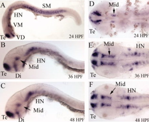
Expression patterns of (A-F) cam-kiin1 by whole mount in situ hybridization. Stages of embryos are indicated in the lower right. At 24 HPF, the cam-kiin1 was predominantly expressed in the telencephalon (T) region, ventral diencephalons (VD), ventral midbrain (VM), and somites (SM). At 36 and 48 HPF, cam-kiin1 was localized in telencephalon (T), diencephalons (Di), midbrain (Mid), and neurons in hindbrain (HN). A-C: Lateral view; D-F: flattened dorsal view. All images are anterior to the left. |
Expression Data
| Genes: | |
|---|---|
| Fish: | |
| Anatomical Terms: | |
| Stage Range: | Prim-5 to Long-pec |
Expression Detail
Antibody Labeling
Phenotype Data
Phenotype Detail
Acknowledgments
This image is the copyrighted work of the attributed author or publisher, and
ZFIN has permission only to display this image to its users.
Additional permissions should be obtained from the applicable author or publisher of the image.
Full text @ Dev. Dyn.