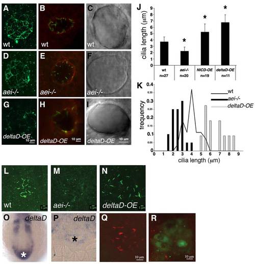
DeltaD regulates cilia length in the Kupffer′s vesicle.(A,D,G) ZO-1 immunostaining of a representative wild-type embryo (A), an aei-/- mutant embryo (D) and a deltaD-overexpressing embryo (G). (B,E,H) aPKC immunostaining of a representative wild-type embryo (B), an aei-/- mutant embryo (E) and a deltaD-overexpressing embryo (H). (C,F,I) DIC images of live embryos showing a wild-type KV (C), a KV from an aei-/- embryo (F) and a KV from a deltaD-overexpressing embryo (I). (J) Quantification of ciliary length at 10-somite stages in wild type, in aei-/- mutants and in embryos where Notch signalling was hyperactivated by overexpressing NICD (NICD-OE) or deltaD (deltaD-OE). Asterisks indicate experimental conditions that produce changes that are significantly different from wild type (P<0.001, Student′s t-test, two tail for two samples assuming unequal variances). (K) Histogram showing the distribution of Kupffer′s vesicle ciliary length at 10-somite stages in aei-/- mutant embryos (black bars), wild-type embryos (black line) and embryos subjected to deltaD-OE (grey bars). (L-N) Confocal images of all z-sections spaced 1 µm apart through the entire ciliated Kupffer′s vesicle at the 10-somite stage. Cilia are labelled with anti-acetylated α-tubulin in a wild-type embryo (L), an aei-/- mutant (M) and an embryo subjected to deltaD-OE (N). (O,P) Wild-type deltaD expression at the 10-somite stage in whole mount (O) and in resin section (P). The white and the black asterisks label the position and the lumen of the Kupffer′s vesicle, respectively. (Q,R) deltaD-MO co-injected with fluorescein lineage tracer in the dorsal forerunner cells (DFCs). (Q) Injected embryo with non-targeted Kupffer′s vesicle cells showing normal length cilia. (R) Fluorescein-positive Kupffer′s vesicle cells with shorter cilia. Error bars indicate s.d.m.
|
