|
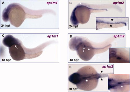
Spatio-temporal expression of ap1m1 and ap1m2. Whole mount in situ hybridization assays were performed on embryos at 24 and 48 hpf. A, C: ap1m1 expression in brain and eye at 24 hpf (A) and 48 hpf (C). Signal is also present at 48 hpf in the gut region (white arrow). B–E: ap1m2 expression. B: ap1m2 is detected at the level of pronephric ducts (black arrowhead) and gut region (white arrow) at 24 hpf. D: White arrowhead indicates digestive tract and white arrow indicates liver at 48 hpf. E: Dorsal view. Black arrowheads indicate the pronephric ducts and white asterisk indicates the intestinal bulb at 30 hpf. All embryos (except E) are mounted lateral view anterior to the left.
|