|
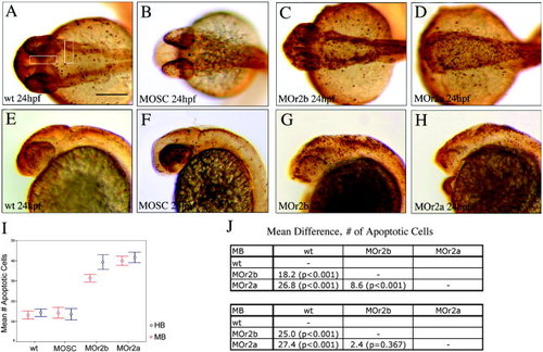
The acvr2a and acvr2b genes mediate apoptosis. A–D: Dorsal views. E–H: Lateral views. Apoptotic cell populations were identified in 24 hours postfertilization acvr2a and acvr2b morpholino oligomer-injected embryos by terminal transferase-mediated dUT nick end-labeling (TUNEL) assay (C, G and D, H, respectively). The number of apoptotic cells in the midbrain and hindbrain of each of 10 wild-type, standard control injected, acvr2a and acvr2b MOs was estimated by counting TUNEL-labeled cells within the white boxed regions (A). I,J: A one-way analysis of variance was performed to quantify differences in apoptotic cell populations.
|