Fig. 4
- ID
- ZDB-FIG-100809-1
- Publication
- Albertson et al., 2005 - Zebrafish acvr2a and acvr2b exhibit distinct roles in craniofacial development
- Other Figures
- All Figure Page
- Back to All Figure Page
|
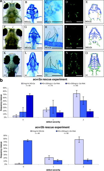
Characterization of acvr2a and acvr2b morphants (MOs). a: Acvr2 morphant cartilages, bones, and teeth. Alcian blue–stained cartilages appeared misshapen and/or missing in acvr2a and acvr2b MOs (B, G) compared with wild-type (wt) controls (L). Quercetin-stained acvr2a and acvr2b MOs revealed abnormal bone development (D and I, respectively) compared with wild-type sibling controls (N). Representative tooth phenotypes are shown (C, H, and M, teeth are outlined in black). The superimposition of bones and cartilages of acvr2a and acvr2b MO and wt embryos reveals distinct defects (E, J, and O, respectively). b: Rescue of morphant phenotypes with coinjected wild-type acvr2a and acvr2b mRNAs. The specificity of the morpholino oligomer phenotypes was confirmed by rescue with wild-type acvr2 mRNAs. Coinjection of wild-type acvr2a mRNA at 200 and 300 ng/μl resulted in rescue of 16% and 67%, respectively. The severity of the defects were classified 0–2, low to high. Coinjection of wild-type acvr2b mRNAs at 200 ng/μl resulted in the rescue of 71% of the acvr2b MO phenotype. Injection of wild-type acvr2b mRNAs at 300 ng/μl resulted in an acvr2b overexpression phenotype in greater than 50% of the injected embryos. Scale bars in a = 200 μm for craniofacial images, 50 μm for enlarged tooth images. |

