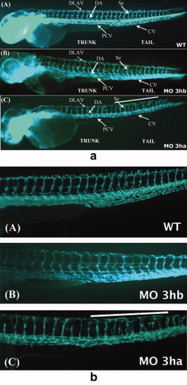
eif3ha morphants also display defects in segmental vessel formation. Panel a: Shown are representative embryos at 3 days postfertilization (dpf) derived from transgenic fli:gfp parents, allowing visualization of the developing vasculature. Panels A and B show the uninjected wild-type (WT) and eif3hb morphant (MO) embryos (6ng/injection), respectively, with normal vasculature pattern, while panel C represents eif3ha morphant embryos with clear defects in segmental vessel formation (70-75%, n ~ 100). Note that the eif3hb morphants received a relatively low dose of morpholino with no apparent brain degeneration phenotype. The caudal vein (CV), posterior cardinal vein (PCV), dorsal aorta (DA), dorsal longitudinal anastomotic vessel (DLAV) are indicated. Panel b: Shown are higher magnification views of the mid-trunk region where irregular segmental vessel formation (Se) is most severe in the eif3ha morphants (panels C) compared with uninjected and eif3hb morpholino injected embryos (panels A and B, respectively). This region in the eif3ha morphants is indicated with a white line in both panel a and panel b.
|
