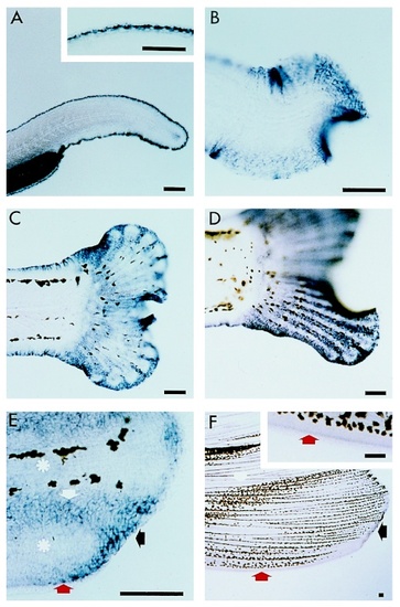
Figure 2. Expression of apoE during caudal fin morphogenesis. Lateral view, anterior to the left. A: 35-hr embryo. ApoE is expressed in the epidermis of the unique median fin fold around the posterior part of the embryo. Inset is a high magnification showing that there is, however, no expression in the most distal layer of cells. B: 7-days, 3 mm, larva. ApoE transcripts become more widely distributed in the epidermis of the tail region. C-E: During larval stages, apoE is expressed in the epidermis surrounding the developing fin rays. C: 21-day, 5.6 mm; D: 24-day, 6.3 mm ; E: 40-day, 10 mm larvae. E: High magnification of two developing fin rays (asterisks) shows that apoE is expressed in the epidermis of the distal part of the fin (black arrow), along the margin of the fin but not in the most external epidermal cell layer (red arrow), and in the inter-ray tissue (white arrow in E). F: ventral lobe of the caudal fin of an adult fish, 33 mm. ApoE is still expressed in the epidermis in the same pattern as during larval development (arrows) but at a much lower level. The inset is a high magnification of the lateral fin margin showing that there is no expression in the most distal epidermal cell layer. Scale bar = 100 m.
|
