
Fig. S2
- ID
- ZDB-FIG-100506-34
- Publication
- Ishimatsu et al., 2010 - Emergence of traveling waves in the zebrafish segmentation clock
- Other Figures
- All Figure Page
- Back to All Figure Page
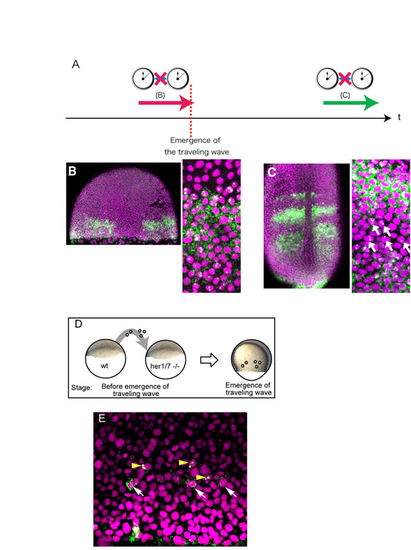
The emergence of the traveling wave does not depend on intercellular communication. (A-C) Inhibition of Notch signaling by DAPT does not affect the emergence of the traveling wave. (A) To examine whether the onset of the her1 traveling wave depends on Notch-dependent intercellular signaling, embryos were treated with the Notch signaling inhibitor DAPT as described previously (Horikawa et al., 2006). (B) An embryo treated with DAPT until the emergence of the traveling wave. The traveling wave was created in the absence of Notch signaling. (C) An embryo treated with DAPT after the emergence of the traveling wave. The spatial pattern of the traveling wave is disturbed, with several transcribing cells detected in the transcription-negative area (arrow). This treatment was performed as a positive control to confirm that DAPT treatment was effective. The two experiments were performed under the same conditions, with the exception of the timing of treatment. Although the horizontal oscillation phase is not exactly the same in the traveling wave at the earlier stage, the occurrence of the traveling wave in the absence of Notch signaling indicates that this wave is not the one relayed from marginal oscillation through Notch signaling. (D,E) We performed transplantation experiments to examine whether unknown intercellular coupling plays a significant role in relaying the marginal her1 oscillation to create the traveling wave. Wild-type cells were transplanted into the her1/7 deletion mutant, which lacks the her1/7 genomic region and thus does not produce any type of her1 oscillation, including the marginal one. If the traveling wave does not depend on intercellular signaling triggered by the margin, the transplanted normal cells can initiate her1 oscillation in the mutant background. (D) Schematic of the cell transplantation assay at the blastula stage. Transplantation was performed at the stage before the onset of her1 marginal oscillation. (E) The transplanted cells show her1 expression with proper phase delay. Since her1/7 mutant cells never express her1, the cells with her1 mRNA signal should be donor cells. The arrowheads indicate cells with her1 transcription, whereas the arrows indicate cells with her1 translation, indicating that the transplanted wild-type cells initiate her1 oscillation with proper phase delay. This result excludes the idea that the traveling wave is triggered by the marginal oscillation through intercellular coupling. Scale bar: 50 μm. |