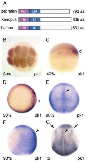
pk1 is expressed maternally and in migrating mesodermal precursors in zebrafish. (A) Homology of Prickle proteins between zebrafish (Pk1), Xenopus (XPk-A) (Wallingford et al., 2002b) and human (BAB71198). The numbers refer to the percentage aminoacid identities between the different orthologues. (B-E) Expression of pk1 at early stages. pk1 is expressed maternally at the eight-cell stage (B, animal view), on the dorsal side at ∼40% epiboly (C, lateral view with dorsal to the right), and around the germ ring at 50% epiboly (D, animal view with dorsal to the right). At 90% epiboly (E, dorsal view, F, lateral view, anterior is up), expression is in dorsal involuted cells and in overlying ectodermal cells and highlighted in the axial cells (arrowheads). At tailbud stage (G, dorsal view, anterior is up), expression is restricted to the presomitic mesoderm, posterior neuroectoderm, the lateral edge of neural plate (arrows) and anterior axial mesodermal cells (black arrowhead), but slightly downregulated in the posterior axial mesodermal cells (red arrowhead). d, dorsal; s, shield.
|
