
Fig. 3
- ID
- ZDB-FIG-100504-46
- Publication
- Chao et al., 2010 - Complexity of cis-regulatory organization of six3a during forebrain and eye development in zebrafish
- Other Figures
- All Figure Page
- Back to All Figure Page
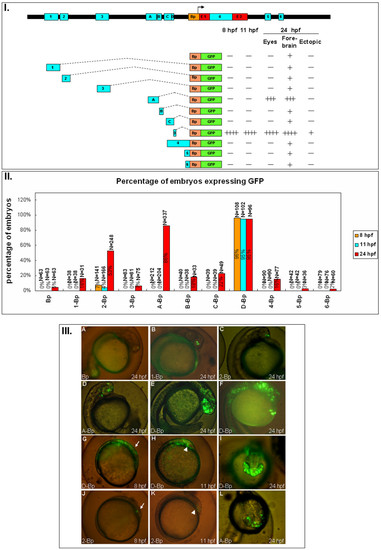
The expression pattern of the ten conserved non-coding modules of zebrafish six3a. I. Different constructs used to study the function of each module. Followed by each construct are the summary of the GFP expression level. Zebrafish embryos injected with each construct expressing GFP at different times (8, 11 and 24 hpf) and regions (eyes, forebrain and ectopic), as in figure 2, the plus and minus indicated the level of GFP intensity. II. The percentages of embryos expressing GFP from different batches are shown with the total numbers of embryos, and three time points: 8 hpf (orange), 11 hpf (blue) and 24 hpf (red). III. Representative GFP expression patterns for each construct in lateral views (A-F, G-H, J-K) or dorsal view (I, L). Most of the images are from 24 hpf except for G and J at 8 hpf, and H and K at 11 hpf (A) Bp-GFP. (B) 1-Bp-GFP. (C) 2-Bp-GFP. (D) A-Bp-GFP. (E) D-Bp-GFP expresses GFP in forebrain and midbrain. (F) D-Bp-GFP expresses GFP extending to the notochord. (G) D-Bp-GFP expression pattern at 8 hpf; the anterior position is marked with an arrow. (H) D-Bp-GFP expression pattern at 11 hpf; the position of the brain is marked with an arrowhead. (I) D-Bp-GFP at 24 hpf showing ventral expression in addition to the forebrain and eye. (J) 2-Bp-GFP expresses at 8 hpf; the anterior position is marked with an arrow. (K) 2-Bp-GFP expression pattern at 11 hpf; the position of the brain is marked with an arrowhead. (L) A-Bp-GFP. |