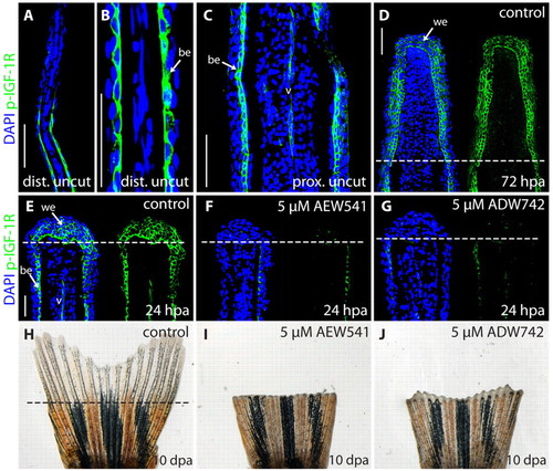
NVP-AEW541 and NVP-ADW742 block Igf1r signaling in zebrafish fins, resulting in inhibition of fin regeneration. (A-G) Longitudinal fin sections stained with phospho-Igf1r antibody (green) and DAPI (blue) to visualize nuclei. (A,B) A distal part of an uncut fin shown at different magnifications. A single cell layer of the basal epidermis labeled with phospho-Igf1r antibody. The distal tip of the fin is devoid of phospho-Igf1r staining. (C) A proximal part of an uncut fin. Phospho-Igf1r antibody marks the basal epidermis and blood vessel (v). (D) A regenerative outgrowth at 72 hpa. The wound epidermis contains phospho-Igf1r-positive cells. The dashed line indicates the amputation plane. (E) A control fin at 24 hpa with phospho-Igf1r-positive cells in the wound epidermis, the basal epidermal layer of the stump, and in the endothelial cells of the blood vessels. Treatment with 5 μM NVP-AEW541 (F) or 5 μM NVP-ADW742 (G) severely reduces phospho-Igf1r staining in the fins at 24 hpa. (H-J) Whole caudal fins at 10 dpa display blocked regeneration after treatment with 5 μM NVP-AEW541 (I) or 5 μM NVP-ADW742 (J), compared with control (H). be, basal epidermis; v, blood vessel; we, wound epidermis. Scale bars: 50 μm in A,C,D,E.
|
