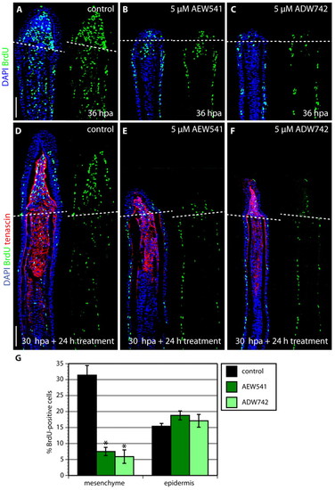
IGF signaling is required for proliferation of blastema cells. (A-C) Longitudinal sections of fins at 36 hpa stained with BrdU antibody (green) and DAPI (blue). Fins treated with either NVP-AEW541 (B) or NVP-ADW742 (C) display decreased mesenchymal proliferation and no blastemal outgrowth, compared with control (A). (D-F) Longitudinal sections of fins after a drug shift: 30 hours at normal conditions followed by 24 hour treatment with 0.05% DMSO (control) (D), 5 μM NVP-AEW541 (E) and 5 μM NVP-ADW742 (F), triply stained with BrdU antibody (green), Tenascin C antibody (red) and DAPI (blue). The dashed line demarcates the amputation plane. In control fins, the blastema displays massive proliferation (D). The smaller size of the outgrowth and reduction of blastemal proliferation demonstrates a block of normally initiated regeneration after the exposure to the drug. (G) The percentage of BrdU-positive cells relative to total number of cells in regenerating fins after DMSO (control) or inhibitor treatment for 24 hours starting at 30 hpa. The counted nuclei were located up to ∼350 μm from the distal tip of the fin sections. Error bars represent the s.e.m. n=6; *P<0.01. Scale bars: 50 μm.
|
