|
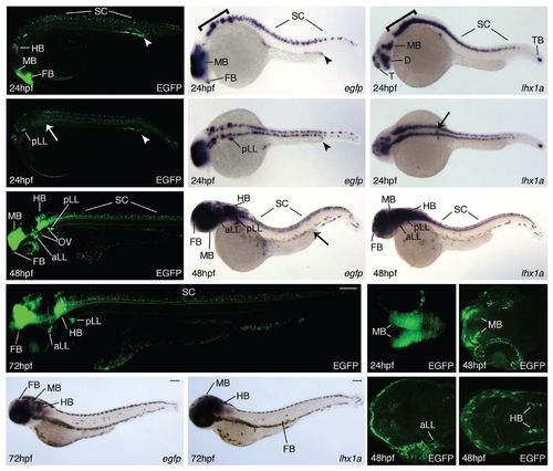
Expression of Tg(lhx1a:EGFP)pt303 during later development.
All animals are oriented such that anterior is to the left. (A-P) Tg(lhx1a:EGFP)pt303 embryos and larvae. ( A , B , G , J , M - P ) Tg(lhx1a:EGFP)pt303 expression. (C,D,H,K) In situ hybridization for egfp. (E,F,I,L) In situ hybridization for lhx1a. (A-F) 24 hpf embryos. Arrow marks position of pronephric proximal tubule and arrowhead delineates distal tubule and duct. (A,C,E) Lateral view, bracket denotes hindbrain. (B,D,F) Dorsal view. (G-I) 48 hpf larvae, arrow in H marks distal tubule and duct in the kidney. (J-L) 72 hpf larvae. (J) Montage of two images to display entire larvae. (M-P) Longitudinal optical sections of transgenic animals. (M) 24hpf embryo. (N-P) 48hpf larva. Abbreviations: aLL, anterior lateral line ganglia; FB, forebrain; HB, hindbrain; MB, midbrain; OV, otic vesicle; pLL, posterior lateral line ganglia; SC, spinal column; TB, tailbud.
|