|
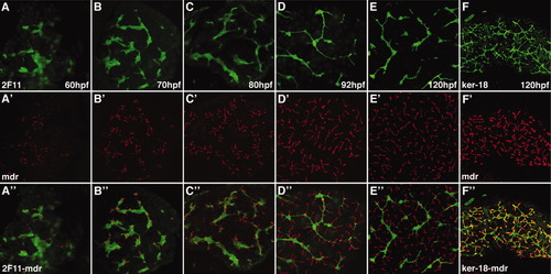
Development of hepatocyte canaliculi and intrahepatic biliary network. Developmental pattern of the 2F11 epitope (A-E) and Mdr epitope, a canalicular transporter (A′-E′) with overlap of the two markers (A″-E″). The length and number of the canaliculi increases between 60 and 120 hpf. The overlap of the two markers shows that each canaliculus develops in close association with the 2F11-positive biliary epithelia. Each canaliculus drains into a single intrahepatic bile duct. The ducts associated with many of the canaliculi in the 120-hpf sample are out of the plane of focus. F, F′, F″: Comparable confocal projections through the liver of a larva immunostained with the keratin-18 and Mdr antibodies. Note overlap (yellow) of the keratin-18 epitope in the terminal ductules with the Mdr protein in the canalicular membrane.
|