
Fig. 3
- ID
- ZDB-FIG-091217-124
- Publication
- Edeling et al., 2009 - Structural requirements for PACSIN/Syndapin operation during zebrafish embryonic notochord development
- Other Figures
- All Figure Page
- Back to All Figure Page
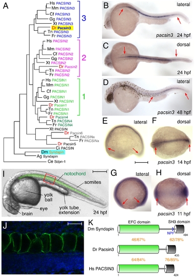
The zebrafish Pacsin3 orthologue. (A) PACSIN family dendrogram (TreeFam accession TF313677). Mm, Mus musculus; Cf, Canis familiaris; Gg, Gallus gallus; Xt, Xenopus tropicalis; Dr Danio rerio; Tn, Tetraodon nigroviridis; Fr, Fugu rubripes; Ci, Ciona intestinalis; Ag, Anopheles gambiae; Ce, Caenorhabditis elegans. (B–H) Embryonic pacsin3 mRNA localization (purple) by whole mount in situ with pacsin3 antisense riboprobe at the various developmental stages noted. Anterior is left. Bar = 250 μm. (I) Lateral view of a 24 hpf control embryo with the notochord pseudocolored in green to highlight the location of this organ. Other structures apparent at this stage are labeled. Bar = 250 μm. (J) Indirect immunolabeling (green) of Pacsin3 with affinity-purified antibodies in the notochord at a lateral region of a fixed 24 hpf embryo, analogous to the red box in I. Nuclei are counterstained with Hoechst (blue). Bar = 50 μm. (K) Schematic of the organizational relatedness and domain structural identity/similarity between selected Syndapin/PACSIN isoforms. |
| Gene: | |
|---|---|
| Fish: | |
| Anatomical Terms: | |
| Stage Range: | 1-4 somites to Long-pec |