
Fig. 4
- ID
- ZDB-FIG-091215-15
- Publication
- Mönnich et al., 2009 - Expression of cohesin and condensin genes during zebrafish development supports a non-proliferative role for cohesin
- Other Figures
- All Figure Page
- Back to All Figure Page
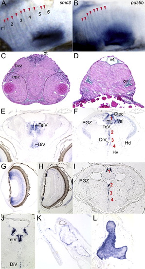
Distribution of cohesin gene expression in embryonic and 2-month-old juvenile brain. (A and B) Close-up of expression of the cohesin subunit smc3 (A) and the associated pds5b (B) at the rhombomere boundaries of 48 hpf zebrafish embryos. The labels r1–6 mark the position of the rhombomeres in (A). Red arrowheads mark the stripes of boundary cells in which the genes are expressed. (C and D) Transverse sections trough the brain of a 48 hpf embryo hybridized with a rad21 riboprobe showing regions of expression in the eye, brain and otic vesicle. (E–G) Transverse sections through brain (E and F) and eye (G) of an 8-week-old juvenile zebrafish hybridized with a rad21 riboprobe. (E) telencephalon; (F) mesencephalon. Mesencephalic proliferation zones are numbered in red 1–4: (1) tectal and torus longitudinalis proliferation zones; (2) diencephalic proliferation zones; (3) dorsal thalamic, posterior tubercular proliferation zones; and (4) hypothalamic proliferation zone. Proliferation zones were identified according to Grandel et al. (2006). (G) Transverse section though eye of 8-week-old zebrafish showing rad21 expression in cells just under the lens and throughout the retina. (H–J) Transverse sections through 8-week-old eye (H) and brain (I and J) hybridized with a pcna riboprobe. (H) Eye showing individual cells in the retina, and cells adjacent to the lens expressing pcna. (I) Mesencephalon; (J) telencephalon. Proliferation zones in (I) numbered 1–4 in red indicate the same regions of proliferation as above, expressing pcna. (K and L) Transverse sections hybridized with a rad21 riboprobe. (K) Scale next to head skeleton. (L) Thymus. bvz, brain ventricular zone; Ctec, tectal commissure; DiV, diencephalic ventricle; epz, eye proliferative zone; Hd, periventricular dorsal hypothalamus; Hv, periventricular ventral hypothalamus; ot, optic tectum; ov, otic vesicle; PGZ, periventricular gray zone of optic tectum; TelV, telencephalic ventricles; TeV, tectal ventricle; TL, torus longitudinalis; v4, 4th ventricle; Val, valvula cerebelli. |
Reprinted from Gene expression patterns : GEP, 9(8), Mönnich, M., Banks, S., Eccles, M., Dickinson, E., and Horsfield, J., Expression of cohesin and condensin genes during zebrafish development supports a non-proliferative role for cohesin, 586-594, Copyright (2009) with permission from Elsevier. Full text @ Gene Expr. Patterns