|
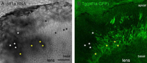
The transition from ptf1a RNA to ptf1a:GFP protein expression. Coronal section through Tg(ptf1a:GFP) embryos with in situ hybridization labelled endogenous ptf1a RNA expression at 48 hpf. (A) Endogenous ptf1a RNA expression as revealed by BM purple visualized in situ hybridization. Labelled cells are found near the apical surface and in the middle of the developing retinal neuroepithelium. (B) Tg(ptf1a:GFP) embryos show a large band of labelled cells in the centre of the neuroepithelium, where differentiating amacrine cells form part of the inner nuclear layer. A few labelled cells are found more apically, where horizontal cells make up the outermost layer of the inner nuclear layer. In some regions apical cells express only the endogenous ptf1a RNA, but not GFP (white asterisks), some co-express both (yellow asterisks), and cells already in the future amacrine layer primarily express only GFP (black asterisks). Scale bar = 20 μm.
|