
Fig. 6
- ID
- ZDB-FIG-090817-31
- Publication
- Moro et al., 2009 - Analysis of beta cell proliferation dynamics in zebrafish
- Other Figures
- All Figure Page
- Back to All Figure Page
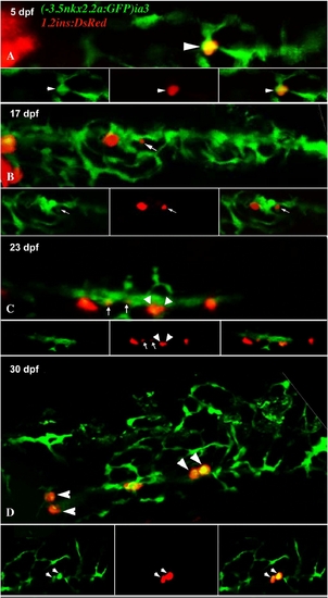
Differentiation of nkx2.2-expressing duct cells into insulin-expressing cells occurs in the intrapancreatic duct during larval stages. (A–D) Projections of confocal stack images at different larval stages of Tg(-3.5nkx2.2a:GFP)ia3/Tg(-1.2ins:dsRed) fish. Single focal plans of channels red, green and their overlay are represented at the bottom of each panel. In panel B the small arrow indicates the budding of an insulin-expressing cell from the duct. (C) Budding of two new insulin-expressing cells (arrows) occurring with a concomitant division of an insulin-expressing cell (arrowheads). (D) Pairs of insulin-expressing cells lie among nkx2.2a-expressing duct cells (arrowheads). |
| Genes: | |
|---|---|
| Fish: | |
| Anatomical Term: | |
| Stage: | Day 5 |
Reprinted from Developmental Biology, 332(2), Moro, E., Gnügge, L., Braghetta, P., Bortolussi, M., and Argenton, F., Analysis of beta cell proliferation dynamics in zebrafish, 299-308, Copyright (2009) with permission from Elsevier. Full text @ Dev. Biol.