
FIGURE
Fig. S3
Fig. S3
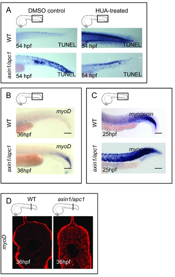
(A) Inhibition of proliferation with HUA resulted in decrease in apoptotic cells in axin1/apc1 embryos. The increase in apoptosis in HUA-treated wild-type embryos, as shown with TUNEL assay, suggests that proliferation has to be tightly regulated. (B) Sustained expression of MyoD+ muscle precursors at 36 hpf, as shown with WISH. Scale bar, 100 μm. (C) Sustained expression of Myogenin+ terminally differentiating cells at 25 hpf, as shown with WISH. Scale bar, 100 μm. (D) Increase in MyoD+ myoblasts at 36hpf, as shown with immunolabelling with anti-MyoD. |
Expression Data
Expression Detail
Antibody Labeling
Phenotype Data
Phenotype Detail
Acknowledgments
This image is the copyrighted work of the attributed author or publisher, and
ZFIN has permission only to display this image to its users.
Additional permissions should be obtained from the applicable author or publisher of the image.
Full text @ PLoS One