
FIGURE
Fig. S3
Fig. S3
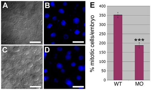
Effect of PrP-1 Knockdown on Apoptosis and Mitosis Rates (A–D) DAPI stainings on 6-hpf embryos (B and D) do not reveal morphological signs of increased apoptosis (such as chromatin condensation and nuclear fragmentation) in detached knockdown cells (C) compared to control embryonic cells (A). (E) PrP-1 morphant embryos show a clear reduction in the number of mitotic cells at 6 hpf (p = 4.3 x 10⁻⁷; triple asterisks [***] indicate statistical significance at p < 0.001); however, neither cell size nor overall cell density seems significantly affected (see main text). Scale bars in (A–D) indicate 10 μm. |
Expression Data
Expression Detail
Antibody Labeling
Phenotype Data
Phenotype Detail
Acknowledgments
This image is the copyrighted work of the attributed author or publisher, and
ZFIN has permission only to display this image to its users.
Additional permissions should be obtained from the applicable author or publisher of the image.
Full text @ PLoS Biol.