
Fig. S1
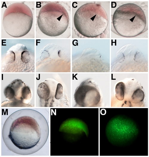
PrP Overexpression Affects Zebrafish Gastrula and Neural Stages (A) Control embryos at the dome stage (4.3 hpf) are morphologically symmetrical and initiate epiboly in a uniform manner. (B–D) Overexpression of zebrafish (zf) PrP-1 ([B] zf PrP-1), PrP-2 ([C] zf PrP-2), or mouse (m) PrP ([D] m PrP) results in earlier morphogenetic movements at one end of the blastodisc, causing a distinctive deformation of the embryo shape (-60%, n = 200, arrowheads). (E–L) At prim-5 (24 hpf) (E–H), and long pec (48 hpf) (I–L) stages, the normal development of the head in control embryos (E and I) contrasts sharply with the small brains and reduced, asymmetric (F–H and J–L), or even fused (K) eyes observed in embryos overexpressing zf PrP-1 (F and J), zf PrP-2 (G and K), or m PrP (H and L). (M–O) The asymmetric epiboly phenotype (M)-seen here for overexpression of zf PrP-1 at 6 hpf- is not due to asymmetric distribution of mRNA, as lateral (N) and animal pole (O) fluorescence views of the same embryo show. (A–D, M, and N) show lateral views; (E–L) rostral views; and (O) animal pole view. |