
FIGURE
Fig. S2
Fig. S2
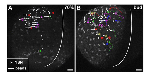
Cortical flow within the YSL is altered in MZoep mutant embryos. (A,B) Trajectories of iYSN (arrowheads) and 0.5 μm diameter fluorescent microspheres (beads; arrows) in the YSL in 70% epiboly (7 hpf; A) and bud stage (10 hpf; B) MZoep mutant embryo. Images are z-projections. Some nuclear and bead trajectories obtained using Motion Tracking Software are shown. Circles indicate the endpoint of each track. White line indicate the dorsal midline of the embryo. Animal is towards the top. Scale bars: 50 μm. |
Expression Data
Expression Detail
Antibody Labeling
Phenotype Data
Phenotype Detail
Acknowledgments
This image is the copyrighted work of the attributed author or publisher, and
ZFIN has permission only to display this image to its users.
Additional permissions should be obtained from the applicable author or publisher of the image.
Full text @ Development