
Fig. 4
- ID
- ZDB-FIG-090310-8
- Publication
- Buckles et al., 2004 - Combinatorial Wnt control of zebrafish midbrain-hindbrain boundary formation
- Other Figures
- All Figure Page
- Back to All Figure Page
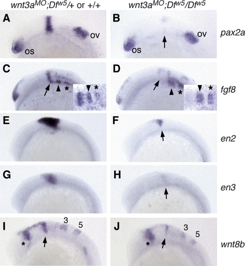
Marker expression in 12-somite stage wnt3aMO;Dfw5/Dfw5 embryos. All panels are lateral views, anterior to the left. The in situ probe used is indicated to the right of each row, and the embryonic genotype is indicated above each column. (A,B) Note the strong reduction in pax2a in wnt3aMO;Dfw5/Dfw5 embryos in the MHB (arrow), while the optic stalk (os) and otic vesicle (ov) expression are unaffected. (C,D) fgf8 is expressed in the MHB (arrows), r2 (arrowheads) and r4 (asterisks). Note specific reduction of MHB fgf8 expression in wnt3aMO;Dfw5/Dfw5 embryos. Insets: dorsal views focused on MHB region, anterior left. (E,F) en2 expression is strongly reduced or absent in wnt3aMO;Dfw5/Dfw5 embryos (arrow) as is en3 (arrow in H). Of note: en3 is not affected in Dfw5/Dfw5 embryos at this stage (Lekven et al., 2003). (I,J) wnt8b is variably reduced in the MHB of wnt3aMO;Dfw5/Dfw5 embryos (arrow), while the forebrain (asterisk), r3 and r5 domains (numbered) are not significantly different from wild type. |
Reprinted from Mechanisms of Development, 121(5), Buckles, G.R., Thorpe, C.J., Ramel, M.C., and Lekven, A.C., Combinatorial Wnt control of zebrafish midbrain-hindbrain boundary formation, 437-447, Copyright (2004) with permission from Elsevier. Full text @ Mech. Dev.