|
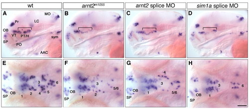
Similar reduction of DA cells in embryos injected with arnt2 or sim1a antisense morpholinos. (A-H) Expression of th at 4 dpf in uninjected wild-type embryos (A,E), arnt2m1055 mutants (B,F), embryos injected with 2.8 ng arnt2 splice morpholino (C,G) and embryos injected with 1.1 ng sim1a splice morpholino (D,H). (A-D) Affected groups are marked by a bracket. (E-H) Magnification of ventral diencephalic groups 1-6. (A-D) Lateral views; (E-H) dorsal views. Anterior towards the left. Scale bar in H: 100 μm for A-D, 50 μm for E-H. Images in E-H represent z-projections from several adjacent focal planes. AAC, arch associated cluster; LC, locus coeruleus; MO, medulla oblongata; OB, olfactory bulb; PO, preoptic region; Pr, pretectum; PT, posterior tuberculum; SP, subpallium; sym, sympathetic CA neurons; VT, ventral thalamic cluster.
|