
FIGURE
Fig. S4
Fig. S4
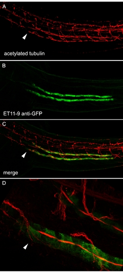
The ET11–9–Positive Segment Marks the Proximal-Most Extent of Multiciliated Cells Two-dpf ET11–9 embryos were stained with anti-acetylated tubulin (A; red) and anti-GFP (B; green). (C) Merge of (A) and (B). (D) Close-up of the anterior (proximal) end of the ET11–9 GFP-positive domain showing compressed, acetylated tubulin-positive bundles of cilia in the lumen of GFP-positive tubule cells. Dorsal acetylated tubulin-positive cells are neurons. Arrows mark the most proximal bundles of cilia originating from pronephric multiciliated cells [23]. |
Expression Data
Expression Detail
Antibody Labeling
Phenotype Data
Phenotype Detail
Acknowledgments
This image is the copyrighted work of the attributed author or publisher, and
ZFIN has permission only to display this image to its users.
Additional permissions should be obtained from the applicable author or publisher of the image.
Full text @ PLoS Biol.