
Fig. 4
- ID
- ZDB-FIG-090220-39
- Publication
- Pei et al., 2009 - Identification of common and unique modifiers of zebrafish midline bifurcation and cyclopia
- Other Figures
- All Figure Page
- Back to All Figure Page
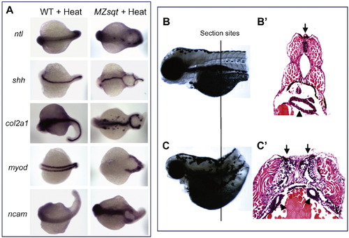
Tissue on both sides of midline bifurcations acquires midline identity. (A) Whole-mount in situ analysis of gene expression. WT and MZsqt embryos were heat treated overnight (22 h) starting at the 64-cell stage and fixed on 1 day post fertilization for whole-mount in situ analysis. All embryos are oriented with a dorsal view, anterior on the left. (B–C) Histological sections of WT and MB embryos. WT and MZsqt embryos were heat treated for 22 h starting at the 64-cell stage and then reared for 2 more days, fixed with 4% paraformaldehyde, paraffin embedded, sectioned (5 μm/section) and stained with hematoxylin and eosin. The approximate section sites for the WT embryo (B) and the MZsqt embryo (C) are indicated. Apostrophes (') indicate the sections from the same specimen. Arrows indicate neural tubes. Arrowheads indicate guts. |
| Genes: | |
|---|---|
| Fish: | |
| Condition: | |
| Anatomical Terms: | |
| Stage: | Prim-5 |
| Fish: | |
|---|---|
| Condition: | |
| Observed In: | |
| Stage: | Prim-5 |
Reprinted from Developmental Biology, 326(1), Pei, W., and Feldman, B., Identification of common and unique modifiers of zebrafish midline bifurcation and cyclopia, 201-211, Copyright (2009) with permission from Elsevier. Full text @ Dev. Biol.