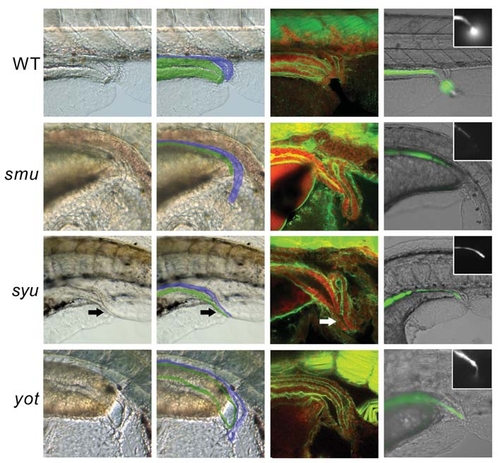
Hh signalling is essential for cloacal development. Bright field images of live Hh pathway mutants and WT siblings (A,B,E,F,I,J,M,N) and confocal scans of embryos stained with α-actin-phalloidin (green) and propidium iodide (red) at 120 hpf (C,G,K,O). Live embryos were injected with fluorescein salt at 72-96 hpf, which accumulates in the gut prior to the opening of the gastrointestinal tract and is then excreted once the gut is functional. Brightfield/fluorescent image merge with the fluorescent channel only shown in the inset (D,H,L,P). The overlay in (B,F,I,N) shows the approximate identity of the relevant tissues, with the posterior gut in green and pronephric ducts in blue. (A,B) WT. By 120 hpf the gastrointestinal tract has fused with the proctodeum and has an independent opening, adjacent to the pronephric ducts. (C) WT. The pronephric ducts have a single narrow opening slightly caudal to the posterior gut. The position where the ducts join is visible in the confocal cross section as an upturned ′U′ shape, with the anterior ducts out of the plane of focus. The posterior gut is surrounded by smooth muscle, and ends with an anal sphincter that is only slightly narrower then the preceding gut. (D) WT embryo excreting fluorescent dye. (E,F) smu. The pronephric ducts are present and are likely to be functional, but the posterior gut is not visible, due to the shape of the embryo and folding of the ventral fins. (G) smu. The pronephric duct caudal to the point where the left and right ducts fuse is much longer in smu mutants because the proctodeum has not opened out. The posterior gut tries to fuse with the proctodeum, but fails, resulting in atresia. (H) smu embryos are completely imperforate (100%, 40/40) and lack peristaltic movement. Application of gentle pressure to the gut moves the dye to the end of the gastrointestinal tract, where it is clear that it is blocked and imperforate. (I,J) syut4 (shha). A channel apparently common between the posterior gut and pronephros (arrow) is suggestive of a fistula between them, but it seems the gut does not fuse correctly with the cloaca leading to imperforate anus. (K) syut4. The posterior gut of syut4 embryos ends blindly dorsal to the proctodeum in most cases. Frequently there is an abortive attempt to fuse with the cloaca leading to the appearance of a fistula between the posterior gut and pronephric duct (arrow). The gastrointestinal tract is completely imperforate – even in instances when there appears to be a fistula. (L) syut4 embryo with imperforate anus/atresia; fluorescent dye can be seen at the blocked end of the gastrointestinal tract in all embryos (100%, 49/49). (M,N) yot (gli2). The pronephric ducts appear to be functional, but are slightly distorted. The gastrointestinal tract is imperforate having failed to fuse with the proctodeum. In this instance peristaltic movements have created a pressure buildup in the distal gastrointestinal tract, causing an outward bulge. (O) yot. The pronephric ducts join together in the correct position, but the usually short single duct appears slightly longer then normal due to the proctodeum failing to open. The posterior gut turns ventrally towards the proctodeum, but fails to fuse with it fully, instead ending blindly adjacent to the pronephric opening. Additionally there is a large reduction in gut girth, probably due to a reduction in smooth muscle in the caudal gut. (P) yot. The majority of yot embryos are imperforate (83%, 15/18), but in the embryos shown here, dye is excreted from the gut through a narrow opening, which is the equivalent of anal stenosis.
|
