
Fig. 1
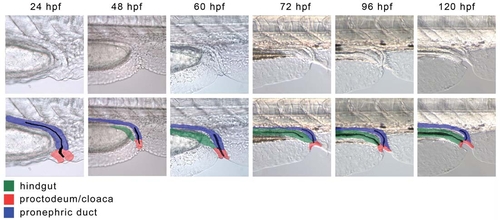
The development of the cloaca in WT embryos. (A-E) Live WT embryos between 24 and 120 hpf. At 24 hpf the proctodeum is visible as a mass of cells at the site of the pronephric opening. The proctodeum surrounds the opening of the pronephric duct at all stages and forms the cloaca. By 48 hpf the posterior gut has reached the cloaca and starts to fuse with it. As the posterior gut becomes canalised from anterior to posterior, the proctodeum invaginates to form the anus. The anal canal, the most distal part of the gastrointestinal tract, is formed by the fusion of the proctodeum with the posterior gut endoderm. By ∼96 hpf the posterior gut lumen is in contact with the ventral edge of the embryo, but is not yet open. At 120 hpf the gastrointestinal tract opens externally, adjacent to the pronephric duct. (G-L) Live WT embryos between 24 and 120 hpf with an overlay showing the identity of various tissues: green, posterior gut; blue, pronephric ducts; red, proctodeum/cloaca. |