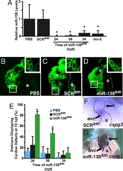
Temporal regulation of miRNA function by antagomiRs in zebrafish. (A) miR-138 RNA levels detected by qRT-PCR in 72-hpf fish embryos treated with antagomiRs from 24, 30, or 34 h postfertilization (hpf) or injected with the 31-nt morpholino (mo-2) compared with PBS- or scrambled antagomiR-treated controls. (B–D) Confocal images of hearts of 72-hpf Tg(myl7:HRAS-mEGFP)s843 transgenic embryos treated with PBS (B), scrambled antagomiR (SCRam) (C), or miR-138 antagomiR (miR-138am) (D) from 24–72 hpf. GFP revealed rounded myocyte morphology (Inset) in miR-138am embryos compared to the normally elongated myocytes seen in controls. (E) Pericardial edema indicating cardiac dysfunction and rounded ventricular myocytes reflecting delayed maturation were observed in varying percentages of 72-hpf embryos treated with PBS, SCRam, or miR-138am from the hpf indicated. (F and G) Ventral view of mRNA in situ hybridization to detect cspg2 expression in hearts of embryos treated with SCRam (F) or miR-138am (G) showed expansion of cspg2 into the ventricle of miR-138am embryos; heart is indicated with dotted lines. Results shown are the average of four experiments in (A) and (E). (v, ventricle; a, atrium; atrioventricular canal (avc); *, P < 0.05.)
|

