
FIGURE
Fig. 2
- ID
- ZDB-FIG-081111-21
- Publication
- Linney et al., 1999 - Transgene expression in zebrafish: a comparison of retroviral-vector and DNA-injection approaches
- Other Figures
- All Figure Page
- Back to All Figure Page
Fig. 2
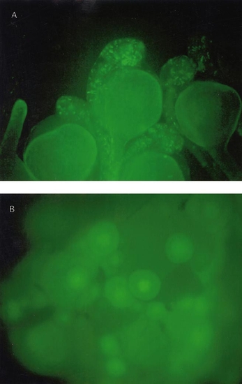
(A) Zebrafish embryos were infected at the 500- to 1000-cell stage with pseudotyped retroviral vector via injection of virions into four separate sites. 24 h after the infection, embryos were examined in the fluorescence microscope for GFP expression. This field is a typical view of the expression obtained. (B) Follicles from an Ef1-α-39-derived adult that was sacrificed to examine the ovaries. Note that the GFP is present in the nucleus (similar observations were made on follicles from a RVEG-19 female). |
Expression Data
Expression Detail
Antibody Labeling
Phenotype Data
Phenotype Detail
Acknowledgments
This image is the copyrighted work of the attributed author or publisher, and
ZFIN has permission only to display this image to its users.
Additional permissions should be obtained from the applicable author or publisher of the image.
Reprinted from Developmental Biology, 213(1), Linney, E., Hardison, N.L., Lonze, B.E., Lyons, S., and DiNapoli, L., Transgene expression in zebrafish: a comparison of retroviral-vector and DNA-injection approaches, 207-216, Copyright (1999) with permission from Elsevier. Full text @ Dev. Biol.