
Fig. S4
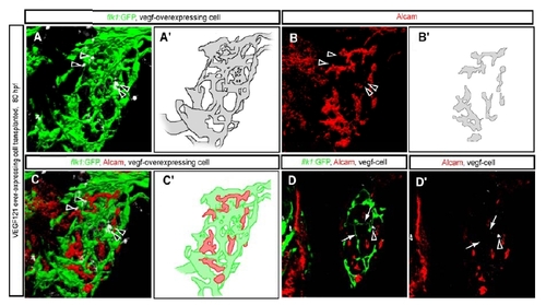
The intrahepatic vascular and biliary networks in larvae containing vegf121 mRNA over-expressing cells in their liver. Tg(flk1:EGFP)s843 larva containing vegf121 mRNA over-expressing cells visualized for GFP (green), Alcam (pseudo-colored red) and donor cell tracer (rhodamine dextran; pseudo-colored white) at 80 hpf. (A) In this transplanted larva, 4 donor cells (arrowheads) located inside the liver disrupt the intrahepatic vascular network (schematically presented in A′). (B) Projected confocal image of the same liver visualized for Alcam expression at 80 hpf. The pattern of the Alcam positive network (schematically presented in B′) reflects the disrupted intrahepatic vascular network. (C) Merged image of Alcam (red) and Tg(flk1r:EGFP)s843 (green) expression shows that the intrahepatic networks still do not intersect. (D) z-plane confocal image of the same larva. Alcam expression and rhodamine dextran are shown separately in D′. Endothelial cells have formed extra vessels (arrows in D and D′) near the vegf121 mRNA over-expressing cells. |