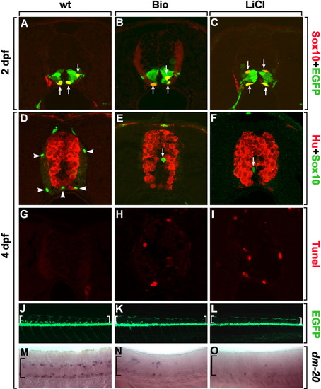
Down-regulation of Wnt signaling is required for the maturation of OPCs. A-I: Transverse sections of spinal cord, dorsal up. A-C: Tg(olig2:egfp) embryos labeled with an anti-Sox10 antibody to detect undifferentiated OPCs at 2 dpf. Arrows indicate Sox10+, olig2:EGFP+ OPCs in control (A), Bio-treated (B), or LiCl-treated (C) embryos from 24-48 hpf. D-F: Combined anti-Hu and anti-Sox10 labeling of control (D), Bio-treated (E), or LiCl-treated (F) embryos from 2-4 dpf. Arrowheads indicate differentiated oligodendrocytes in the white matter (D) and arrows indicate undifferentiated OPCs near the ventricle (E, F). G-I: TUNEL staining of control (G), Bio-treated (H), or LiCl-treated (I) embryos from 2-4 dpf. J-L: Side views of Tg(olig2:egfp) embryos, dorsal to the top and anterior to the left. J: Control embryo has numerous olig2:EGFP+ oligodendrocytes in the dorsal spinal cord (brackets). Bio-treated (K) and LiCl-treated (L) embryos from 2 dpf show only a few olig2:EGFP+ cells in the dorsal spinal cord (brackets) at 4 dpf. M-O: Lateral views of embryos hybridized with dm-20 RNA probes, which label mature oligodendrocytes at 4 dpf. M: Control embryos have many dm-20+ mature oligodendrocytes in the spinal cord (bracket). Bio-treated (N) and LiCl-treated (O) embryos have few cells marked by dm-20, suggesting that OPCs fail to differentiate into mature oligodendrocytes (brackets).
|
