
Fig. 8
- ID
- ZDB-FIG-081013-53
- Publication
- Lele et al., 1999 - Disruption of zebrafish somite development by pharmacologic inhibition of hsp90
- Other Figures
- All Figure Page
- Back to All Figure Page
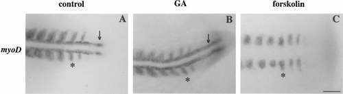
Adaxial cells form in GA-treated but not in 14-h-old forskolin-treated embryos. (A) Control. (B) GA. (C) Forskolin. Adaxial cells are demonstrated based on their early, strong expression of myoD. Adaxial cells (arrow) could be detected directly adjacent to the notochord prior to somitogenesis in both control and GA-treated embryos but did not form in embryos treated with forskolin. In contrast, the myoD-expressing lateral presomitic cells which will go on to form fast muscle fibers were normal in all three groups of embryos (asterisks in A–C). Treatment with GA and forskolin was initiated at 50% epiboly. Scale bar, 50 μm. |
Reprinted from Developmental Biology, 210(1), Lele, Z., Hartson, S.D., Martin, C.C., Whitesell, L., Matts, R.L., and Krone, P.H., Disruption of zebrafish somite development by pharmacologic inhibition of hsp90, 56-70, Copyright (1999) with permission from Elsevier. Full text @ Dev. Biol.