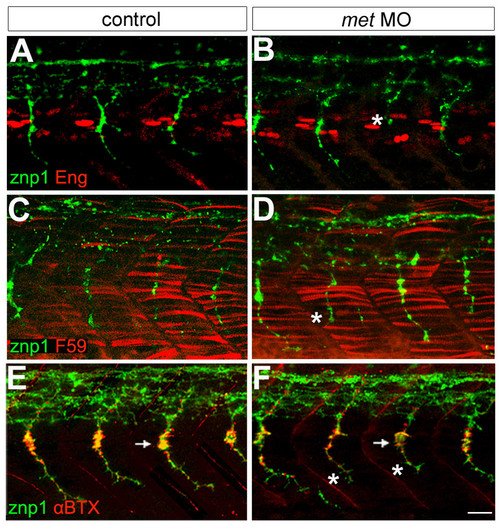
Met appears unnecessary for muscle and neuromuscular junction formation. (a,b) Engrailed antibody (Eng, red) labeling showing muscle pioneer cells and znp1 antibody labeling showing motor axons (green). In met MO-injected embryos, some CaP axons are truncated (asterisk). (c,d) F59 antibody (red) labeling showing fast muscle fibers and znp1 antibody staining showing motor axons (green). F59 labeling appears the same in control (c) and met MO-injected (d) embryos, which have some truncated CaPs (asterisk). (e-f) αBTX (red) labeling showing AChRs and znp1 antibody labeling showing motor axons (green). The distribution of AChRs appears the same in control (e) and met MO-injected embryos (f) that have some truncated CaP axons (asterisks); however, it appears that the number of AChRs may be decreased at the myoseptal varicosity (arrows) by MO injection. For each experiment, 8 spinal hemisegments plus somites were examined in each of 21–33 met MO-injected embryos and 8 spinal hemisegments plus somites in each of 15 controls. Scale bar, 20 μm.
|
