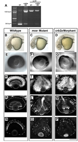
Injection of crb2a splice-blocking morpholinos phenocopies the moe mutations. (A) crb2a splicing, measured by RT-PCR, is reduced by injection of splice-blocking morpholino antisense oligonucleotides. Morpholino injection results in the amplification of a 1097 bp fragment. The reactions were also performed with no reverse transcriptase (no RT). (B-D) Head morphology of 30-hour wild-type (B), moeb476 mutant (C) and crb2a morphant (D) embryos. (E-G) The retinal pigmented epithelium is uniform in wild type (E) and patchy in moeb781 mutants (F) and crb2a morpholinos-injected embryos (G) at 48 hours. (H-J) The three nuclear layers are clearly defined in the wild-type retina (H), but are not defined in the moeb781 mutant (I) and the crb2a morphant (J) at 3 days. (K-M) Retinal ganglion cells (labeled using Zn5 antibodies) localize to the innermost part of the retina in the wild-type retina (K), but are disorganized and scattered throughout the retina in moeb781 mutants (L) and crb2a morphants (M) at 3 days. (N-P) Rod photoreceptors (visualized by genetically-encoded GFP) localize to the outermost part of the retina in the wild-type retina (N), but are disorganized and scattered throughout the retina in moeb781 mutants (O) and crb2a morphants (P) at 4 days. Eyes are outlined. Scale bars: in D, 500 μm for B-D; in P, 100 μm for E-P.
|
