
Fig. 3
- ID
- ZDB-FIG-080702-28
- Publication
- Lee et al., 2008 - Zebrafish blowout provides genetic evidence for Patched1-mediated negative regulation of Hedgehog signaling within the proximal optic vesicle of the vertebrate eye
- Other Figures
- All Figure Page
- Back to All Figure Page
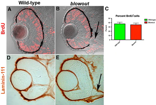
Retinal cell proliferation and Bruch’s membrane formation are both normal in blowout. (A, B) Wild-type (A) and blw (B) embryos were exposed to BrdU from 42 hpf to 48 hpf and immediately sacrificed. BrdU positive cells (red) are observed throughout the retinas of wild-type and blw mutant embryos in similar proportions (C; no statistical difference—Fisher’s exact test). Additional exposure periods (24–36 hpf, 72–96 hpf) resulted in identical regions of proliferation in the retinas of wild-type and blw mutants (data not shown). (D, E) Bruch’s membrane, a basement membrane at the posterior of the eye, highly expresses the laminin-111 protein (Lee et al., 2007). Shown here are images of 12 μm cryosections from wild-type (D) and blw (E) whole-mount embryos stained for laminin-111 protein at 48 hpf. Laminin-111 levels and distribution are similar between wild-type and blw embryos. The optic stalk appears abnormal in blw, as it remains connected to the retina at 48 hpf (arrows in panels B, E), while in wild-type siblings it has degenerated. Transverse sections, dorsal is up in all images. |
| Fish: | |
|---|---|
| Condition: | |
| Observed In: | |
| Stage: | Long-pec |
Reprinted from Developmental Biology, 319(1), Lee, J., Willer, J.R., Willer, G.B., Smith, K., Gregg, R.G., and Gross, J.M., Zebrafish blowout provides genetic evidence for Patched1-mediated negative regulation of Hedgehog signaling within the proximal optic vesicle of the vertebrate eye, 10-22, Copyright (2008) with permission from Elsevier. Full text @ Dev. Biol.