
Fig. 2
- ID
- ZDB-FIG-080702-27
- Publication
- Lee et al., 2008 - Zebrafish blowout provides genetic evidence for Patched1-mediated negative regulation of Hedgehog signaling within the proximal optic vesicle of the vertebrate eye
- Other Figures
- All Figure Page
- Back to All Figure Page
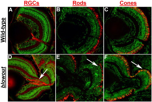
Retinal development is largely normal in blowout. Immunohistochemical analysis of retinal ganglion cells (RGCs) via zn8 staining (A, D), rod photoreceptors via zpr3 staining (B, E) and red/green double cones via zpr1 staining (C, F) in transverse retinal cryosections. All retinal cell types are present and are properly distributed in blw mutants, and retinal cell numbers are also similar between wild-type and blw mutant eyes. RGC organization is affected in blw mutants where RGC axons do not assemble into a tight bundle as they exit the eye (arrow in panel D). Photoreceptor outer segments are present in the retinal territories that are not contained within the eye cup (arrows in panels E, F). Antibody stains are red and nuclei are counterstained green with Sytox Green. Dorsal is up in all images. |
| Antibodies: | |
|---|---|
| Fish: | |
| Anatomical Terms: | |
| Stage: | Day 5 |
| Fish: | |
|---|---|
| Observed In: | |
| Stage: | Day 5 |
Reprinted from Developmental Biology, 319(1), Lee, J., Willer, J.R., Willer, G.B., Smith, K., Gregg, R.G., and Gross, J.M., Zebrafish blowout provides genetic evidence for Patched1-mediated negative regulation of Hedgehog signaling within the proximal optic vesicle of the vertebrate eye, 10-22, Copyright (2008) with permission from Elsevier. Full text @ Dev. Biol.