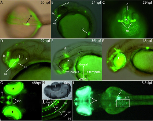
GFP expression pattern in the stable P0-pax6b:gfp transgenic zebrafish. (A) Dorsal view of a 20 hpf transgenic embryo showing GFP expression in the developing pancreas. (B, C) Lateral and frontal views at 24 and 29 hpf showing GFP expression in the ventral telencephalon, the dorsal diencephalon and the optic stalks. (D, E, F) Lateral views of an eye of 29 hpf, 36 hpf and 48 hpf transgenic embryos. GFP expression starts at about 29 hpf in a small cluster of cells in the ventronasal retina, near the optic stalks, and subsequently spreads to the entire retina (white arrows). (G) After 2 days of development, expression is observed in the neuroretina, the optic nerves as well as the ventral telencephalon and some cells of the diencephalon. (H, H′) At 10 dpf, expression is clearly detected in several layers of the retina, mainly the inner plexiform layer, the inner nuclear layer, the outer plexiform layer and the outer nuclear layer. (I) Finally, from 3.5 dpf, the transgenes are expressed in the enteroendocrine cells of the intestine and in some neurones of the mesencephalon. Abbreviations: d, diencephalon; e, eye; ent, enteroendocrine cells; gcl, ganglion cell layer; hb, hindbrain; inl, inner nuclear layer, ipl, inner plexiform layer; l, lens; mb, midbrain; nt, neural tube; oc, optic choroid; on, optic nerves; onl, outer nuclear layer; opl, outer plexiform layer; os, optic stalks; p, pancreas; pcl, photoreceptor cell layer; r, retina; rpe, retinal pigmented epithelium; tc, telencephalon. A, G and I are dorsal views (anterior to the left); B, D, E and F are lateral views (anterior to the left); H, H′ are cross sections in the eye of a 10 dpf embryo.
|
