
Fig. 4
- ID
- ZDB-FIG-080604-40
- Publication
- Chi et al., 2008 - Genetic and Physiologic Dissection of the Vertebrate Cardiac Conduction System
- Other Figures
- All Figure Page
- Back to All Figure Page
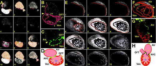
Development of the Fast Specialized Cardiac Conduction Tissue (A and E) Sequential calcium activation images of a 100 hpf (A) Tg(cmlc2:gCaMP)s878 heart and a 21 dpf (E) Tg(cmlc2:gCaMP)s878 ventricle during a single cycle in a live zebrafish. (B and F) The 100 hpf (B) and 21 dpf (F) optical maps of calcium excitation represented by isochronal lines every 60 ms. Ventricular calcium activation initiates along trabeculae at 100 hpf and 21 dpf and breaks through the rest of the myocardium propagating from OC to base at 100 hpf and apex to base at 21 dpf. (C and G) Confocal images of the heart at 100 hpf and 21 dpf. (C) The 100 hpf Tg(flk1:EGFP)s843 (green) larvae stained with rhodamine phalloidin (red) reveal that the hearts have completed cardiac looping and that the ventricles have formed trabeculae.(G) At 21 dpf, the ventricle forms a distinct apex. (D and H) Schematic representation of the hearts shown in (C) and (G). Numbers indicate temporal sequence of calcium activation in the heart. Yellow arrows indicate direction of cardiac conduction. Green circles indicate slow conduction nodes, and blue circles indicate fast conduction pathway. Atr, atrium; Ven, ventricle; AVC, AV canal; Tr, trabeculae. |
| Gene: | |
|---|---|
| Fish: | |
| Anatomical Terms: | |
| Stage Range: | Day 4 to Days 21-29 |
| Fish: | |
|---|---|
| Observed In: | |
| Stage Range: | Day 4 to Days 21-29 |