
Fig. 2
- ID
- ZDB-FIG-080604-2
- Publication
- Postel et al., 2008 - Zebrafish integrin-linked kinase is required in skeletal muscles for strengthening the integrin-ECM adhesion complex
- Other Figures
- All Figure Page
- Back to All Figure Page
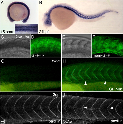
Ilk–GFP cellular localization. (A,B) Whole mount ISH with dig-labelled antisense ilk mRNA at 15-somite stage (A and inset) and 24 hpf (B). Wt embryos injected with synthetic mRNA encoding GFP–ILK (C and D) or memGFP (E and F). Images were taken at the 10-somite stage (15 hpf) at the region of the forming somites. The GFP–Ilk protein shows a predominant cytoplasmic localization at this stage. (G,H) Uninjected wt embryo (G) and a wt embryo injected with synthetic mRNA encoding GFP–Ilk (H) at 24 hpf. GFP–Ilk protein localization at the somite boundaries is indicated by arrowheads. (I,J) Anti-paxillin antibody staining and confocal images of a wt embryo (I) or loc/ilk mutant embryo (J) at 3 dpf. |
| Genes: | |
|---|---|
| Antibody: | |
| Fish: | |
| Anatomical Terms: | |
| Stage Range: | 10-13 somites to Protruding-mouth |
Reprinted from Developmental Biology, 318(1), Postel, R., Vakeel, P., Topczewski, J., Knöll, R., and Bakkers, J., Zebrafish integrin-linked kinase is required in skeletal muscles for strengthening the integrin-ECM adhesion complex, 92-101, Copyright (2008) with permission from Elsevier. Full text @ Dev. Biol.