
FIGURE
Fig. 8
Fig. 8
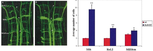
Supernumerary reticulospinal neurons are present in desb420 mutants. Dorsal view of a confocal image (anterior to the top) of a RMO44 labeled wild-type (A) and desb420 mutant (B) embryo. Somata of affected cells are labeled in (A) and arrowheads point to their corresponding axons. Note the increased number of axons in the mutant embryo. (C) Cell counts of affected reticulospinal neurons. Cells on both sides of the midline were analyzed for wild-type (n = 14) and mutant (n = 14) embryos. See Fig. 7 legend for details regarding statistics. Mth, Mauthner cell. Scale bar, 20 μm. |
Expression Data
Expression Detail
Antibody Labeling
Phenotype Data
| Fish: | |
|---|---|
| Observed In: | |
| Stage: | Long-pec |
Phenotype Detail
Acknowledgments
This image is the copyrighted work of the attributed author or publisher, and
ZFIN has permission only to display this image to its users.
Additional permissions should be obtained from the applicable author or publisher of the image.
Reprinted from Developmental Biology, 237(2), Gray, M., Moens, C.B., Amacher, S.L., Eisen, J.S., and Beattie, C.E., Zebrafish deadly seven functions in neurogenesis, 306-323, Copyright (2001) with permission from Elsevier. Full text @ Dev. Biol.