FIGURE
Fig. 2
- ID
- ZDB-FIG-080424-44
- Publication
- Novince et al., 2003 - Cadherin expression in the inner ear of developing zebrafish
- Other Figures
- All Figure Page
- Back to All Figure Page
Fig. 2
|
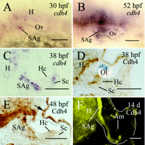
Expression of cadherin-4 message (cdh4) (A–C), and Cadherin-4 immunoreactivity (Cdh4) (D–F) in the inner ear of developing zebrafish. Panels (A) and (B) are lateral views of the otic vesicle of whole-mount embryos. Dorsal is up and anterior is to the left. Panels (C–F) are cross sections of the otic vesicle. The intense labeling of the epidermal tissue in panel (E) (arrow) has been shown to be non-specific, by preabsorption studies (Liu et al., 2001b). Panel (F) is from a section adjacent to panel (F) in Fig. 1. Ol, otolith; other abbreviations are the same as in Fig. 1. Scale bar: 50 μm. |
Expression Data
Expression Detail
Antibody Labeling
Phenotype Data
Phenotype Detail
Acknowledgments
This image is the copyrighted work of the attributed author or publisher, and
ZFIN has permission only to display this image to its users.
Additional permissions should be obtained from the applicable author or publisher of the image.
Reprinted from Gene expression patterns : GEP, 3(3), Novince, Z.M., Azodi, E., Marrs, J.A., Raymond, P.A., and Liu, Q., Cadherin expression in the inner ear of developing zebrafish, 337-339, Copyright (2003) with permission from Elsevier. Full text @ Gene Expr. Patterns

